Phantom chest
The prototype vest is calibrated against calf lungs in a liquid mixture to imitate the dielectric properties of the chest.
Our vest provides a diagnostic Point Of Care Test using MWI for the early diagnosis of PE.
Explore nowPE is a life-threatening condition in which a blood clot - an embolus originating and travelling from a thrombosed deep vein (DVT) blocks one or more of the pulmonary arteries impairing the oxygenation of the blood by the lungs. Symptoms include sudden breathing difficulty, coughing up blood, and chest pain. Larger emboli can potentially cause strain on the right ventricle due to resistance to pumping blood and could potentially result in a fatal, sudden cardiac arrest.
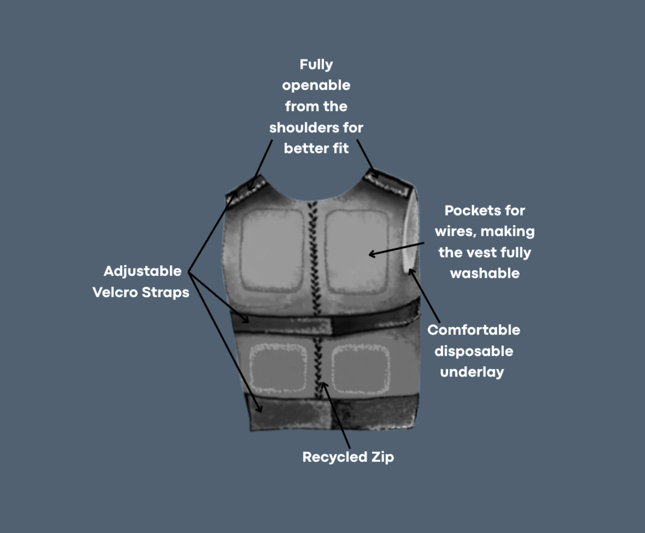
We’ve developed a lightweight, portable microwave imaging vest as a fast, affordable alternative to costly CTPA scans for diagnosing pulmonary embolism. The vest serves as a simple point-of-care (POCT) device that can allow for early, accessible detection of Pulmonary Embolism (PE) without ionising radiation or delays - this would ultimately improve outcomes for both patients and doctors, revolutionsing lung imaging. In emergency settings, where every minute is crucial, increasing the speed of diagnoses would turn lost time into saved lives. As well as this, missed or delayed diagnoses can be fatal; in primary care settings, atypical presentations of PE are often missed - our device would aim to combat this issue, offering GPs a more advanced diagnostic tool kit to make referrals easier.
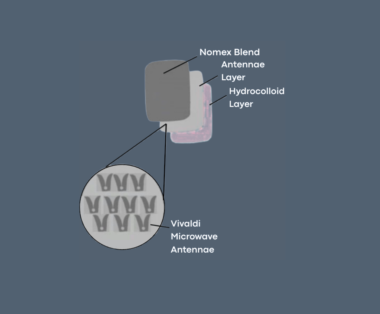
Microwaves emitted by a set of antennae placed around the vest are scattered due to the tissue’s dielectric properties. Clot tissue, oedema and lack of perfusion - localised signs of PE - all lead to distinctive changes in the dielectric properties of the parenchyma. The Vector Network Analyser (VNA) gives microwave absorption, transmission and reflection values, which are then used to calculate the tissue's dielectric constant via an AI based software. These values are used to construct the scattering matrix. Colour-coded images are formed from data by tomographic reconstruction algorithms with comparison to a ‘phantom’ model of healthy lungs. AI based algorithms with Machine Learning detects the presence and location of the clot.
The prototype vest is calibrated against calf lungs in a liquid mixture to imitate the dielectric properties of the chest.
A large random sample of consenting hospital patients with and without PE are used to test the accuracy. The results are compared to a CTPA scan, and clinicians label the blood clot on the MWI. The large data set is used to train a ML algorithm
The ML algorithm is tested on unfamiliar scans, using samples with a lower, the same or higher PE proportion to guarantee accuracy. If the vest has an accuracy of above 95% and passes statistical analysis - it is ready for the next trial.
A&E and GP practices are given the device which is used to test on suspected PE - with healthcare professionals providing feedback. If the MWI scans consistently align with the CTPA hospital scans, the device is ready to be used
The device must be certified by the Medicines and Healthcare products Regulatory Agency (MHRA) and and an endorsement from NICE Medical Technologies Evaluation Programme (MTEP) could streamline device adoption. Introducing it to NHS Health Innovation Network (HIN) would help with integration
The device is listed on the market after company registration. Bulk deals could be secured with private buyers, e.g. airports. Deals are signed with ICBs to sell the devices at affordable prices to GPs and care homes - mediated by the PCNs. At the same time, compatible software is added to the NHS for seamless integration (28)